June 29, 2023
According to the Missouri Radiation Control Program (MRCP)—an entity of the Missouri Department of Health and Senior Services—as of May 2023 there are close to 5,000 facilities in Missouri with ionizing radiation equipment, and 2,106 of those are dental sites. (1) All x-ray units must undergo radiation safety testing, and the scheduling depends on what type of equipment a dental practitioner has installed in their office. This inspection must be performed by an approved Qualified Expert (QE), individuals the MRCP has recognized and approved as having a significant background and training in radiation physics and safety, as well as proficiency in surveying devices producing ionizing radiation. In this article, dental site requirements from the MRCP, general survey information and safe radiation practices will be discussed.
Registration Dates vs Survey Dates
All facilities that have operational ionizing radiation equipment must complete registration documents from the state every two years and update any important information. The survey (inspection) timeline depends on the equipment.
All intraoral wall units, handheld/portable units and panoramic units are considered a class D and are required to be tested every six years prior to their due date; all cone-beam computed tomography units (CBCT 3D) are class E and are required to be inspected every three years due to the higher radiation output and dose. A practice that has both a CBCT unit and other dental x-ray units will have equipment on both survey schedules. (2)
The MRCP sends reminders for both the registration and survey dates to all practices generally 90 days before they are due, and the MDA works with the MRCP to provide an updated list of facilities and due dates at modental.org/radiationinspections.
Noncompliance with either the registration renewal or the required radiation safety survey will result in a warning, followed by a cease-and-desist mandate. When a dental practice moves locations or adds an x-ray unit to its site, there are additional stipulations from the MRCP. If the office relocates, even if it is just down the street, all equipment in the new location must be surveyed. In addition, when a unit is added to a practice, it must be inspected. If the unit is a handheld, panoramic or wall unit, it must be surveyed within 90 days of installation; if it is a CBCT unit, it must be inspected within 30 days. Should a dentist set up a new practice, all x-ray units must be surveyed for acceptance testing and registered with the MRCP. (2) QEs help dental sites navigate the compliance process with adherence to radiation safety.

Figure 1: Partial setup of an intraoral unit.
What are we really inspecting and looking for during a survey?
QEs are testing a variety of important aspects of the dental x-ray units that all boil down to important radiation safety basics: ensuring the dose to the patient is as safe and as low as possible; the output of the unit is operating safely within set parameters; and, the operator of the equipment and surrounding staff will be safe from leakage and scattered radiation as well. In addition, we look at how the unit is set for making exposures to the patient and make recommendations if needed for patient safety.
General radiation physics aspects of the dental x-ray machine survey include testing the kVp, mA, timer, exposure rate, half value layer, reproducibility, linearity, and general mechanics of the dental x-ray unit itself (see Fig. 1). When panoramic and cone-beam CT units are surveyed, scattered radiation surveys will also be performed in the surrounding areas. CBCT units will generally produce at least 10 times as much scattered radiation compared to panoramic units. (3) Some CBCT manufacturers involve additional quality control testing protocols using phantoms, such as resolution capabilities, uniformity, collimation alignment and evaluation of materials, air and water.
For a quick physics refresher, kilovoltage (kV) is the energy and penetration power of the x-ray beam; milliamperage (mA) and time (in seconds) together represent mAs, which regulates the amount of x-ray photons being produced. If you double the mAs, you double the exposure rate and therefore the dose to the patient. When these values are out of limits, the unit must be serviced and recalibrated and sometimes replaced. After the testing is complete, the updated summary results form is submitted to the MRCP by the QE.

Figure 2: External rectangular collimators for intraoral units. Image courtesy of XDR Radiology.
How Much Radiation Dose Is the Patient Really Receiving?
The answer to that question depends on the type of equipment producing the radiation, technique used and receptor type. With most dentists using digital sensors instead of film, the dose to the patient is lower than in previous decades and can be reduced 40 to 70 percent with digital imaging. (3) However, the digital world allows for a wider exposure latitude regarding radiation output, meaning the operator can use a variety of technical factors and still have an acceptable image. The set factors chosen might produce more or less radiation than necessary, as the sensor and digital processing will adjust to produce an image. This is not the case with the old-style film imaging, when doubling the output resulted in an image that was twice as dark. It is important the radiation output be routinely surveyed by the QE to ensure the technical factors are within acceptable limits.
In addition, the National Council on Radiation Protection and Measurements (NCRP) recommends utilizing rectangular collimation for intraoral radiographs, which is usually an attachment to the round cone end of the unit (see Fig. 2). This practice improves image quality and reduces patient dose by four to five times; the beam size will more closely match the image receptor shape. The operator does need to be more precise in the exposure alignment preparation so anatomy cutoff will not occur on the image, and it is noted that this technique is not currently in use in most offices. (3) For those sites still using film, higher speed film, such as E or F speed, must be utilized to decrease patient dose while still maintaining image quality. (3)
We are all exposed to background radiation, including cosmic radiation, every day. Professional radiology organizations, such as the Radiological Society of North America and the American College of Radiology, compare the medical dose to patients to being exposed to background radiation for a better understanding of exposure dose. For example, a series of intraoral x-rays taken on a patient is equivalent to about one day of background radiation; a panoramic x-ray is equivalent to about three days of background radiation; and, a cone-beam CT scan is equivalent to about 22 days of background radiation.4 Many of the CBCT units have parameter selections that include normal and also higher definition outputs, which can significantly increase the patient’s dose. Comparatively, a chest x-ray dose is about 10 days of background radiation, and a chest CT scan is about two years’ worth. (4)
There are risks associated with all x-ray imaging, and the benefit to the patient must outweigh the risk. Ionizing radiation can cause damage to DNA and increases a chance of developing cancer. (3)
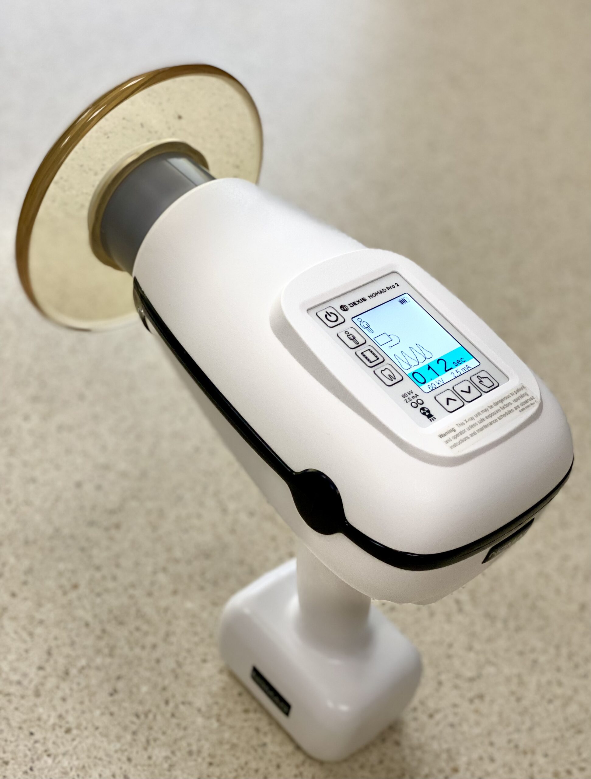
Figure 3: Example of an approved handheld dental x-ray unit.
Are Handheld X-Ray Units Safe?
There are currently seven manufacturers approved by the MRCP for handheld dental x-ray devices.5 These x-ray units are increasing in popularity due to their ease of use by being lightweight and portable in multiple operatories within a practice. The output is similar to an intraoral wall unit, with additional built-in housing shielding for operator safety, and the attached leaded acrylic scatter shield must remain in place during exposure to protect the operator (3,5) (see Fig. 3).
The NCRP states the operator of a handheld x-ray unit is not required to wear a protective lead apron although the state of Missouri recommends one be worn.3,5 It is also advised the facility provide personal dosimeters to operators of these devices for the first year of use to determine whether ongoing monitoring is needed. Personal dosimeters should be worn for occupational workers who are likely to receive an annual effective dose of 100 mrem or higher (1 mSv). (3,6)
What About Shielding the Patient?
According to the NCRP’s Report No. 177, “Technological and procedural improvements have eliminated the requirement for the radiation protective apron; however, some patients expect it and may request it. Thyroid shielding is required for patients due to the high radiosensitivity of the thyroid when it will not interfere with the exam.” A significant portion of the dose to the body is from internal scattered radiation which cannot be shielded. (3) The MRCP supports these statements.
There are several organizations that have current campaigns to lower patient dose and improve radiation safety. Image Gently is an alliance that advocates lowering the exposure factors and using a smaller field of view for pediatric imaging. (7) Image Wisely aims to educate imaging professionals on using appropriate techniques and radiation dose monitoring for safety in adult imaging.8 In summary, there are multiple methods for dental practices to utilize to keep patients and staff safe while performing radiographic imaging.
Intraoral, panoramic and cone-beam CT imaging all have an excellent purpose for specific anatomical diagnosis and procedures. With proper technique, shielding and routine testing being employed, QEs assist sites with radiation safety equipment surveys to ensure these units continue to be operated with adherence to standardized limits and keeping dose as low as is reasonably achievable for the safety of all patients and dental staff.
Stephanie Patrick, BS, RT (R)(M)(QM) is a Qualified Expert with Heldebrandt Consulting and travels throughout Missouri performing radiation safety inspections for sites including dental settings. She has been in the radiologic sciences with experience in radiation physics and safety for more than 30 years and enjoys providing educational radiology information to facilities. Heldebrandt Consulting is an MDA Solutions Center Allied Business Participant. Click to visit their Solution Center listing. Images are property of the author except where noted. This article also appeared in the Focus MDA 2023 Summer issue.
References
- Missouri Department of Health and Senior Services, Bureau of Diagnostic Service, Missouri Radiation Control Program. (2023). Radiation Control. Radiation Control, Health & Senior Services. Retrieved April 29, 2023, https://health.mo.gov/safety/radprotection/
- Missouri Department of Health and Senior Services, Bureau of Diagnostic Service, Missouri Radiation Control Program. (2017, November). Guidance for dental facilities use of QEs – Missouri. Retrieved April 27, 2023, https://health.mo.gov/safety/radprotection/pdf/Guidance-for-Dental-Facilities-Use-of-QEs.pdf
- National Council on Radiation Protection and Measurements. (2019, December 19). Radiation Protection in Dentistry and Oral & Maxillofacial Imaging: Recommendations of the National Council on Radiation Protection and Measurements. NCRP Report No. 177.
- Radiological Society of North America (RSNA) and American College of Radiology (ACR). (2022, November 1). Radiation Dose. Radiologyinfo.org. Retrieved April 29, 2023, https://www.radiologyinfo.org/en/info/safety-xray
- Missouri Department of Health and Senior Services, Bureau of Diagnostic Service, Missouri Radiation Control Program. (2021, September 14). Usage of handheld dental x-ray units in the State of Missouri. Retrieved April 29, 2023, https://health.mo.gov/safety/radprotection/pdf/usage-of-hand-held-xray-units.pdf
- Missouri Department of Health and Senior Services, Bureau of Diagnostic Service, Missouri Radiation Control Program. (2015, August). Guidance for dental facilities, radiation badges and shielding. Retrieved April 29, 2023, https://health.mo.gov/safety/radprotection/pdf/Guidance-for-Dental-Facilities-Radiation-badges-and-shielding.pdf
- Image Gently Alliance. (2014). Image Gently During Dental Procedures. imagegently. Retrieved April 30, 2023, http://www.imagegently.org/Procedures/Dental
- American College of Radiology. (2023). Image Wisely. Retrieved April 30, 2023, https://www.imagewisely.org/About-Us
- 19 CSR 20-10, Code of State Regulations. (2003, January 29). Rules of Department of Health and Senior Services, Division 20 – Division of Environmental Health and Epidemiology, Chapter 10 – Protection Against Ionizing Radiation. Retrieved April 29, 2023, https://s1.sos.mo.gov/cmsimages/adrules/csr/current/19csr/19c20-10.pdf
- Center for Devices and Radiological Health. (2015, September 18). Radiation safety considerations for X-ray equipment for handheld use. U.S. Food and Drug Administration. Retrieved April 29, 2023, https://www.fda.gov/regulatory-information/search-fda-guidance-documents/radiation-safety-considerations-x-ray-equipment-designed-hand-held-use
- Center for Devices and Radiological Health. (2023, February 21). Medical X-ray imaging. U.S. Food and Drug Administration. Retrieved April 29, 2023, https://www.fda.gov/radiation-emitting-products/medical-imaging/medical-x-ray-imaging
